• Welcome to BirdForum, the internet's largest birding community with thousands of members from all over the world. The forums are dedicated to wild birds, birding, binoculars and equipment and all that goes with it.

    Please register for an account to take part in the discussions in the forum, post your pictures in the gallery and more.
ZEISS DTI thermal imaging cameras. For more discoveries at night, and during the day.

New specimen of Palaeeudyptes (1 Viewer)

albertonykus

Well-known member
Xia, B.-Y., J.-L. Pei, and Q.-G. Li (2024)
A new penguin fossil from Seymour Island and reassessment of taxonomy and diversity of Eocene Antarctic penguins
Palaeoworld (advance online publication)
doi: 10.1016/j.palwor.2024.04.007

Eocene penguins from Seymour Island play an important role in studies related to the taxonomy and evolution of the Sphenisciformes stem group. Among these penguins, the Palaeeudyptes species are particularly noteworthy for their unusually large size and the contentious nature of their classification criteria. In this study, we describe a new penguin skeleton with a well-preserved tarsometatarsus discovered in the Upper Eocene of Seymour Island, Antarctica. The new fossil exhibits tarsometatarsal characteristics of Palaeeudyptes but differs from two species of Palaeeudyptes previously found on Seymour Island, providing insights on the morphological diversity and evolutionary history of early penguins. We conduct normality and unimodality tests on Palaeeudyptes taxa from Seymour Island to reassess the hypothesis that size differences between the two species of this genus could be attributed to sexual dimorphism in a single species. The results revealed that size differences are unlikely due to sexual dimorphism. We also use the linear discriminant analysis to evaluate the taxonomic criteria for the two Palaeeudyptes species discovered in the Antarctic region. The data showed an overlap in the size distribution, indicating weakness in the classification criteria. Reassessing previous samples and establishing an additional diagnosis based on critical anatomical features could potentially resolve this issue.
 
Systematic palaeontology

Class Aves Linnaeus, 1758
Order Sphenisciformes Sharpe, 1891
Family Spheniscidae Bonaparte, 1831

Genus Palaeeudyptes Huxley, 1859

Type species: Palaeeudyptes antarcticus Huxley, 1859.

Included species: P. antarcticus Huxley, 1859, P. gunnari Wiman, 1905, P. marplesi Brodkorb, 1963, P. klekowskii Myrcha, Tatur and Del Valle, 1990

Palaeeudyptes sp

Material:
CUGB P2003, a partial skeleton of a single individual, comprising a nearly complete tarsometatarsus in addition to fragmentary forelimb (a right ulna, a left radius, and a manual phalanx) and hindlimb (femurs, tibiotarsi, and pedal phalanges) bones.

Occurrence: Seymour Island, Antarctic Peninsula; Submeseta Formation

Referral to the genus Palaeeudyptes: The specimen exhibits a distinctive combination of tarsometatarsal features, closely resembling Palaeeudyptes and distinguished from other Eocene penguins on Seymour Island: the tarsometatarsus is large and robust (distinguishing in small-bodied Sphenisciformes: Archaeospheniscus Delphinornis, Marambiornis, Marambiornopsis, and Mesetaornis); the tarsometatarsus is smaller than in Anthropornis and falls within the range of Palaeeudyptes (as indicated in Table 1); the medial margin is clearly concave without
any pronounced convexity (unlike in Anthropornis); the medial hypotarsal crest is undivided (subdivided in Anthropornis); the medial proximal vascular foramen is smaller and more proximal compared to its lateral counterpart (as in Palaeeudyptes; opposite to Anthropornis); the lateral dorsal longitudinal sulcus is deep (as in Palaeeudyptes)
Furthermore, this specimen presents a few tarsometatarsal features that deviate from previous reports on Seymour Island Palaeeudyptes. The plantar opening of the medial proximal vascular foramen is located within the hypotarsal sulcus, separated from the medial margin by the medial hypotarsal crest (unlike its placement
somewhat more medially relative to the medial hypotarsal crest or distal to the medial hypotarsal crest in Palaeeudyptes). The tuberosity for insertion of the cranial tibial muscle is less marked than in Palaeeudyptes gunnari. The lateral margin is slightly concave, with the trochlea moderately laterally deflected.

Fred


Fig. 1. Articulated skeleton CUGB P2003, including a nearly complete tarsometatarsus, wing elements (ulna, radius, and manual phalanx) and leg elements (femurs, tibiotarsi, and pedal phalanges).
1715590288157.png


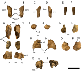
Fig. 2. Wing and leg elements of CUGB P2003 from the Submeseta Formation of Seymour Island. (A, B) Right ulna; (A) dorsal view; (B) ventral view. (C, D) Left radius; (C) dorsal view; (D) ventral view. (E, F) Manual phalanx; (E) dorsal view; (F) ventral view. (G, H) Right femur; (G) cranial view; (H) caudal view. (I, J) Femoral head of the left femur; (I) proximal view; (J) distal view. (K, L) Medial condyle of the left femur; (K) caudal view; (L) medial view. (M–P) Distal end of the left tibiotarsus; (M) cranial view; (N) caudal view; (O) medial view; (P) lateral view. (Q, R) Proximal end of the right tibiotarsus; (Q) cranial view; (R) caudal view. Abbreviations: aaf, antitrochanter articular face; cam, caudal margin; ccc, cranial cnemial crest; cil,
cranial intermuscular line; clf, capital ligament fossa; crm, cranial margin; ec, extensor canal; fms, fibular medial sulcus; ft, fibular trochlea; iom, interosseus margin; is, intercnemial sulcus; lc, lateral condyle; lcc, lateral cnemial crest; mc, medial condyle; osb, ossified supratendinal bridge; ps, patellar sulcus; udc, ulna dorsal condyle; uvc, ulna ventral condyle. Scale bar: 50 mm.
1715590317527.png


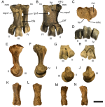
Fig. 3. Foot elements of CUGB P2003 from the Submeseta Formation of Seymour Island. (A–F) Left tarsometatarsus; (A) dorsal view; (B) plantar view; (C) proximal view; (D) distal view; (E) medial view; (F) lateral view. (G–J) Distal end of the right tarsometatarsus; (G) dorsal view; (H) plantar view; (I) medial view; (J) lateral view.
(K–N) Pedal phalanges; (K, M) dorsal view; (L, N) plantar view. Abbreviations: II, trochlea II; III, trochlea III; IV, trochlea IV; hs, hypotarsal sulcus; ldls, lateral dorsal longitudinal sulcus; lm, lateral margin; lpvf, lateral proximal vascular foramen; mc, medial cotyle; mhc, medial hypotarsal crest; mm, medial margin; mpvf, medial
proximal vascular foramen; psf, plantar supratrochlear fossa. Scale bar: 20 mm.
1715590658404.png
 
Last edited:

Users who are viewing this thread

Back
Top